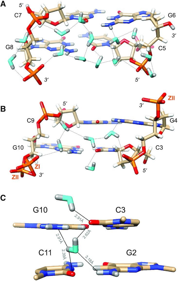
Figure 3.

Water structure in the minor groove. Transverse hydration patterns that link cytosine O2 keto oxygens and guanine N2 amino groups to backbone phosphates. (A) Base pairs C5:G8 and G6:C7 and (B) base pairs C3:G10 and C4:C9. Water oxygen and deuterium atoms are colored in cyan and gray, respectively, and DNA deuterium and hydrogen atoms are colored in gray and white, respectively. H-bonds (D…O) are drawn as thin solid lines, and strand polarities are indicated. ZI and ZII refer to two different phosphate orientations that are characterized by distinct ϵ and ζ backbone torsion angles. (C) The sole example of a water molecule bridging O2 keto oxygens of adjacent cytosines in the minor groove. Electrostatically favorable interactions by this water are not limited to O2 (cytosine) but also involve N2 (guanine), i.e. the lone electron pair of N2(G10) and one of the deuterium atoms of N2(G2). D…N and D…O distances are indicated by thin solid lines.
