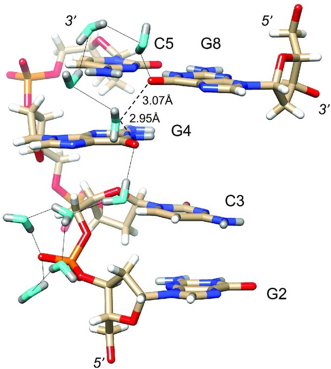
Figure 5.

Longitudinal hydration pattern linking O6 keto oxygen of G and backbone phosphates on one side of the convex surface. Water oxygen and deuterium atoms are colored in cyan and gray, respectively, and DNA deuterium and hydrogen atoms are colored in gray and white, respectively. H-bonds (D…O) are drawn as thin solid lines. Note the formation of pentagons involving four water molecules and either a phosphate oxygen or an O6 keto oxygen. A water molecule positioned between G4 and G8 lies within H-bonding distance from O6 keto oxygens (dashed lines). However, its particular orientation, i.e. both deuterium atoms are directed away from O6 atoms, renders H-bond formation unlikely.
