Figure 1.

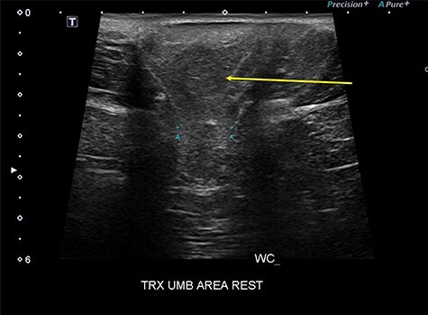
Abdominal wall ultrasound showing the one parietal peritoneal lipoma in the hernia sac (yellow arrow) and the 2 cm hernia neck (A-A).

Abdominal wall ultrasound showing the one parietal peritoneal lipoma in the hernia sac (yellow arrow) and the 2 cm hernia neck (A-A).