Fig. 3.

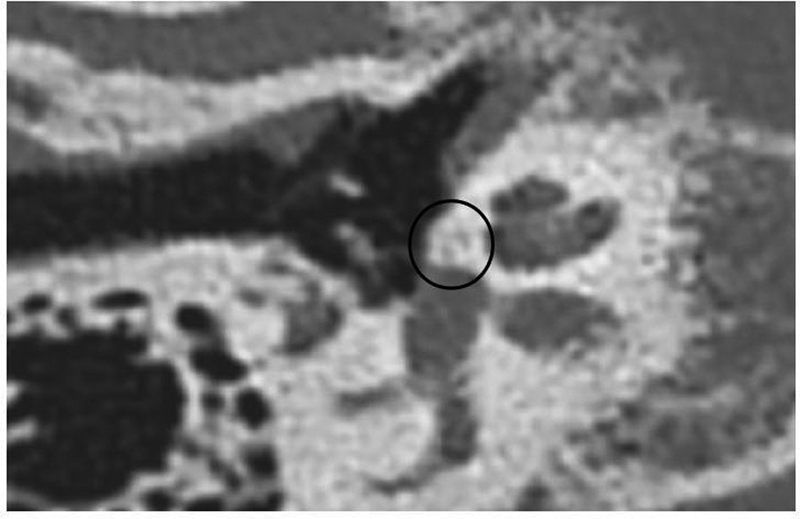
Computed tomography scan of temporal bone. Axial view at the level of the oval window, showing the presence of contour irregularities on the surface of the otic capsule (black circle), typical of inactive otosclerosis. Source: the authors.

Computed tomography scan of temporal bone. Axial view at the level of the oval window, showing the presence of contour irregularities on the surface of the otic capsule (black circle), typical of inactive otosclerosis. Source: the authors.