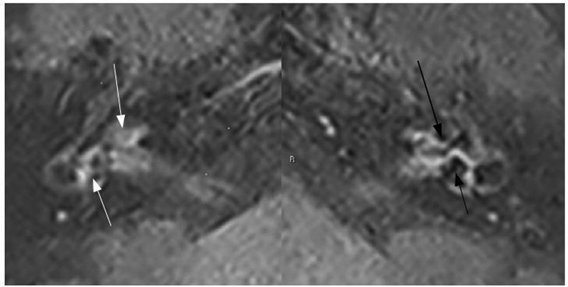
Fig. 8.

Magnetic resonance imaging of the brain, 4 hours after double injection of gadolinium, at the level of the membranous labyrinth, REAL IR sequence, axial view. Areas with signal voids on the left ear might be observed, corresponding to the cochlea and vestibule, compatible with endolymphatic hydrops (black arrows). The contralateral inner ear has a comparative normal appearance (white arrows). Source: the authors.
