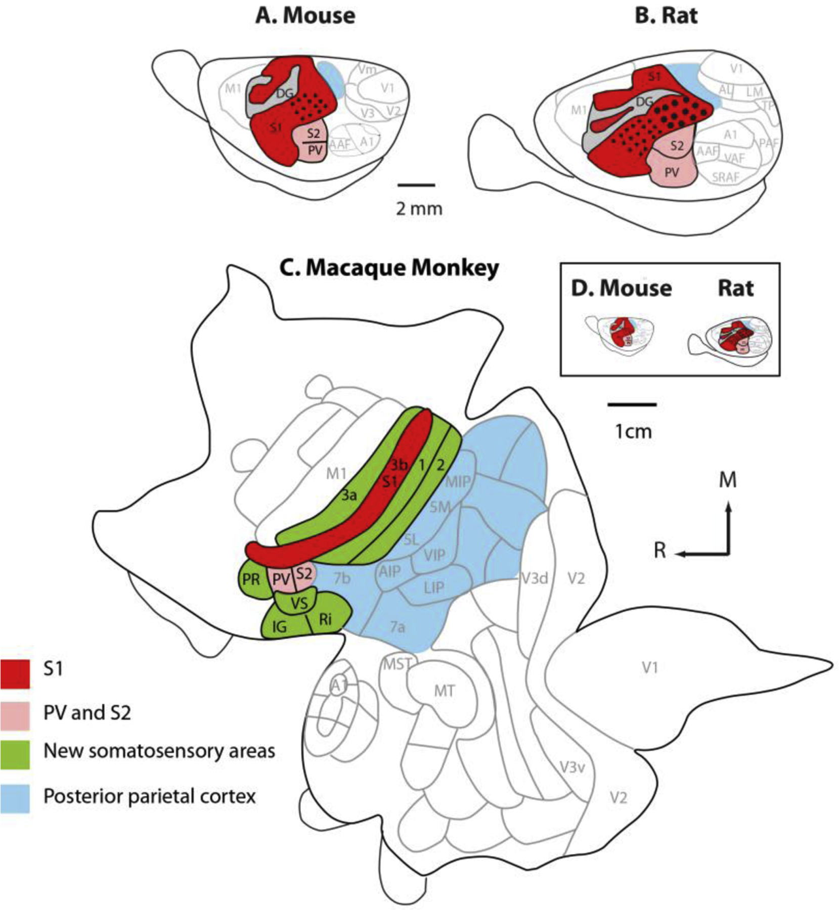
Figure 4. The organization of somatosensory cortex in mice, rats and, macaque monkeys.

All three species have a primary somatosensory area (red), a second somatosensory area, and a parietal ventral area (pink). In macaque monkeys, additional areas that process somatosensory inputs have emerged over the course of evolution (green). Posterior parietal cortex has greatly expanded and includes multiple cortical fields in macaque monkeys. When mice and rat cortices are drawn to scale (D) the enormous difference in the size of the cortical sheet in these rodent models and macaque monkeys is striking. Adapted from Dooley and Krubitzer, 2013.
