Fig. 3.
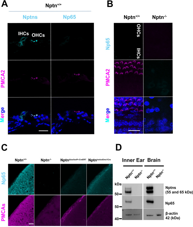
Np65 expressed in the inner ear is not detectable in hair cells. A Mid-modiolar cochlear sections from Nptn+/+ mice were labeled with DAPI and antibodies against PMCA2 and against neuroplastin 55 and 65 (Nptns, left) or specific for Np65 (Np65, right). Neuroplastin was detected in inner (IHCs) and outer hair cells (OHCs), but Np65 was not detected. Scale bar = 30 µm. B Cochlear whole mounts of Nptn+/+ and Nptn−/− mice were labeled with DAPI, antibodies against PMCA2, and antibodies specific for Np65. Np65 could not be detected in inner (IHC) or outer hair cells (OHC). Scale bar = 20 µm. C Sections from the AC of Nptn+/+, Nptn−/−, NptnΔlox/loxPrCreERT, and Nptnlox/loxEmx1Cre mice were labeled with antibodies specific for Np65 and antibodies detecting all PMCAs. Np65 was detected in the AC of Nptn+/+ and in reduced quantity of Nptnlox/loxEmx1Cre mice, but not in Nptn−/− and NptnΔlox/loxPrCreERT mice. Notice that the expression of PMCAs depends on the expression of Np. Scale bar = 30 µm. D Western blot analysis of expression of neuroplastin and Np65 in the brain and the inner ear. Antibodies against neuroplastin 55 and 65 (Nptns) and specific for Np65 (Np65) detected the respective isoforms in the brain and the inner ear of Nptn+/+ but not of Nptn−/− mice

