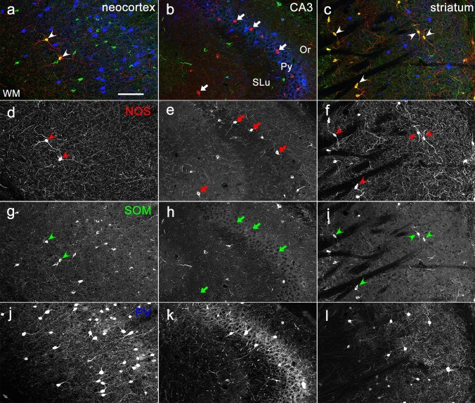
Fig. 1.
NOS immunoreactivity in three different brain regions. Each section showing the neocortex (a, d, g, j), CA3 region of the hippocampus (b, e, h, k) and striatum (c, f, i, l) was obtained from the same animal. Pseudo-color images in (a–c), consisting of immunolabeling for NOS (red), SOM (green), and PV (blue), are shown separately in (d–f), (g–i), (j–l), respectively. Arrows and arrowheads indicate NOS-positive and NOS/SOM-double positive neurons, respectively. Or, stratum oriens; Py, stratum pyramidale; SLu, stratum lucidum; WM, white matter. Scale bar = 100 µm

