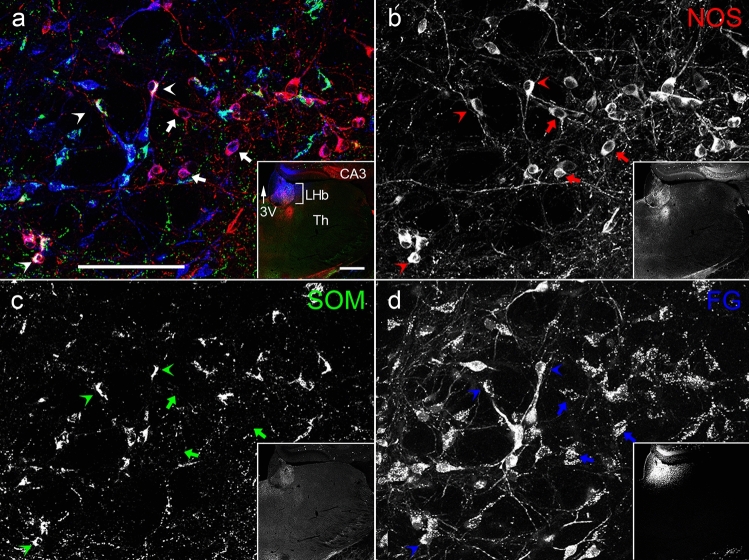
Fig. 7.
The colocalization relationship between retrogradely labeled FG, which was injected into the LHb, and NOS and/or SOM in the EPN. The pseudocolor images in (a) and Inset consist of NOS (red), SOM (green), and FG (blue) immunoreactivities, which are shown separately in (b–d). NOS/FG double-positive neurons and NOS/SOM/FG triple-positive neurons are shown by arrows and arrowheads, respectively. 3V, third ventricle; CA3, CA3 region of the hippocampus proper; Th, Thalamus. The scale bars in (a) and Inset are 100 and 500 µm

