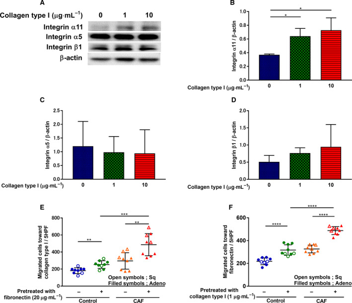
Fig. 5.

Collagen type I‐mediated integrin subunit regulation and fibroblast migration. After HFL‐1 were cultured and incubated with different doses of human collagen type I for 48 h, (A) proteins were extracted and subjected to western blot analysis to detect (B) ITGA11, (C) integrin α5, and (D) integrin β1 expression levels. (B, C, D; n ≥ 3, unpaired Student's t‐test) Vertical axis: expression of protein normalized to β‐actin. (E) After preincubation with human fibronectin (20 µg·mL−1) for 8 h, CAFs‐ and control fibroblasts‐mediated migration toward collagen type I (1 µg·mL−1) was measured. Vertical axis: number of migrated cells per 5HPF. (F) After preincubation with collagen type I (1 µg·mL−1) for 48 h, CAFs‐ and control fibroblasts‐mediated migration toward fibronectin (20 µg·mL−1) was measured. (E, F; n = 9, unpaired Student's t‐test) Vertical axis: number of migrated cells per 5HPF. Each symbol represents one patient. Filled symbols represent lung adenocarcinoma. Open symbols represent squamous cell lung cancer. The values represent the mean ± SD. *P < 0.05 **P < 0.01, ***P < 0.001, ****P < 0.0001.
