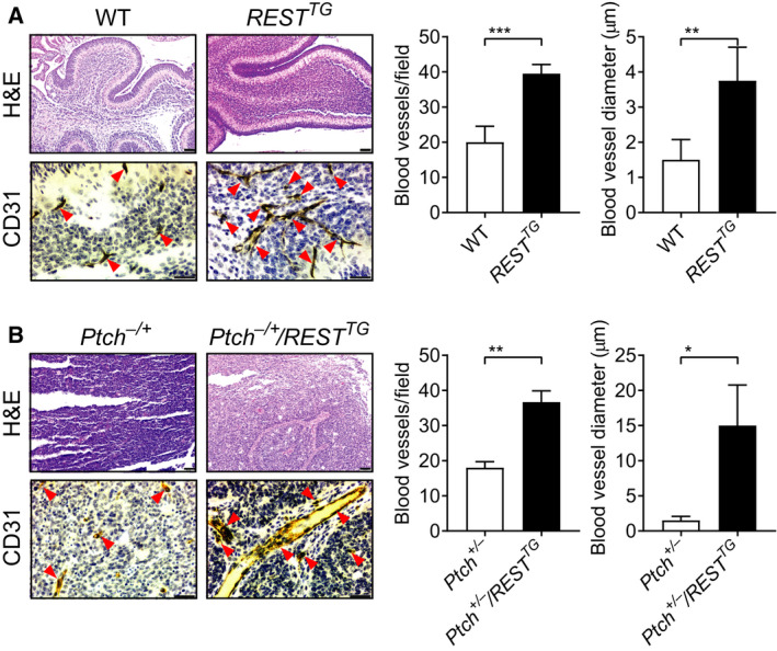
Fig. 1.

REST promotes vasculature in RESTTG cerebella and Ptch+/−/RESTTG tumors. H&E staining and IHC for CD31 were performed on (A) cerebellar sections from WT and RESTTG mice and (B) tumor sections from Ptch+/− and Ptch+/−/RESTTG transgenic mice, to demonstrate the vasculature changes (left panels). Arrowheads show the blood vessels. Quantitation of blood vessels in sections (n = 3; three fields /section), and average blood vessel diameter is shown in the right panels for A & B. Scale bars in A, B; H&E = 20 μm; CD31 = 10 μm. Data show individual variability and means ± SD. P‐values were obtained using Student's t‐test. *P < 0.05, **P < 0.01, ***P < 0.001.
