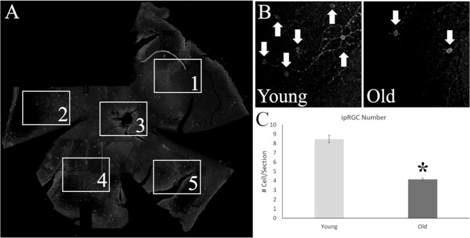
Fig. 5. Histological analysis of intrinsically photosensitive retinal ganglion cells (ipRGC) from the retinas of old and young mice stained for melanopsin.
A Whole mount retinas of the mice were divided into four petals, one image for counting was taken for each petal and from a central location. B The number of total melanopsin positive cells (White Arrows) were lower in old mice (Right Panel) than in their younger counterparts (Left Panel). C Statistical analysis of the number of cells per section demonstrated a significant difference between the age groups, with a 50% reduction in number of cells in older mice. *p < 0.05.

