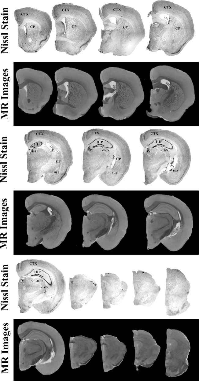
Fig. 6. High-resolution T1 weighted magnetic resonance images compared to Nissl stained brain sections from one representative mouse.
Structures observed within the stained section are clearly identified within the scans. These structures include the cortex (CTX), caudate nucleus (CP), hippocampus (HIP), habenula (EPI), basolateral amygdala complex (BLA), optic tract (opt), superior colliculus (SC), and lateral geniculate nucleus (LGN). The greater resolution (32 µm) utilized in this manuscript allowed for the identification of subregion of the LGN, ventral (vLGN) and dorsal (dLGN) components.

