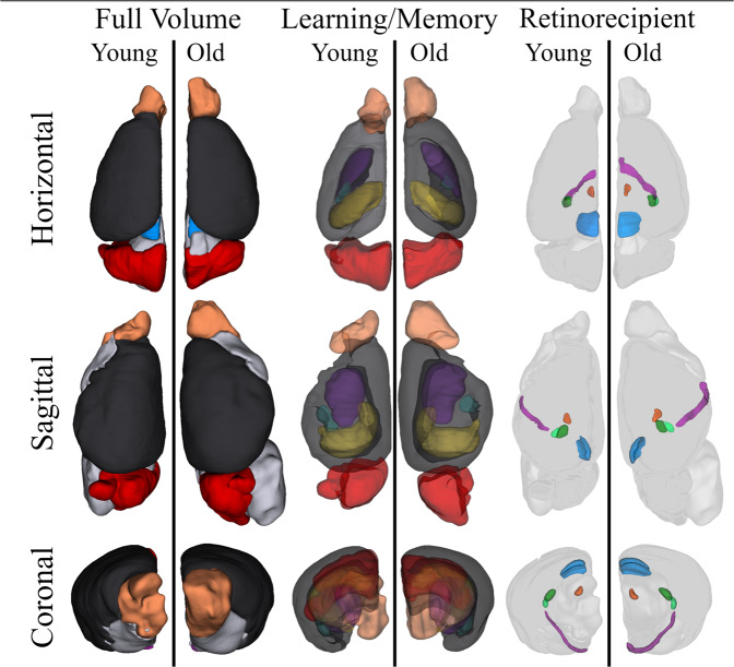
Fig. 7. Three-dimensional volume rendering of learning/memory and retinorecipient non-image forming visual regions in young and old mice.
Volumes for the cortex (black), olfactory bulb (light orange), hippocampus (yellow), amygdala (teal), caudate nucleus (dark purple), cerebellum (red), superior colliculus (blue), geniculate complex (dorsal, dark green; ventral, light green), habenula (dark orange) and the optic tract (light purple) are shown in three orientations. The brains of the two age groups are represented in identical proportions to demonstrate the relative size differences between visual structures. Most notably, a greatly enlarged optic tract is observed within older mice.

