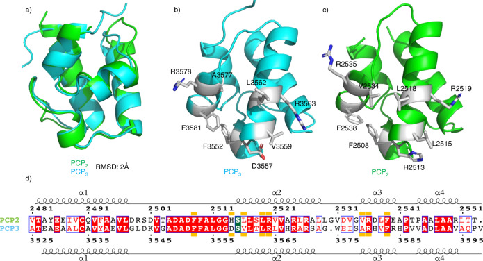
Fig. 3. Comparison of PCP2 and PCP3 domains from fuscachelin biosynthesis.
a Structural alignment of PCP2 and PCP3 domains (RMSD 2 Å). b Crystal structure of the PCP3 domain (PDB ID 7KW3) showing the position of side chains for interaction with C-domain based on PCP2. c Crystal structure of the PCP2 domain showing side chains interacting with the C-domain. d Sequence alignment of both PCP2 and PCP3 domains with the C domain interface indicated by orange blocks (conserved residues highlighted in red, similar residues shown in red text); site of posttranslational modification highlighted in green.

