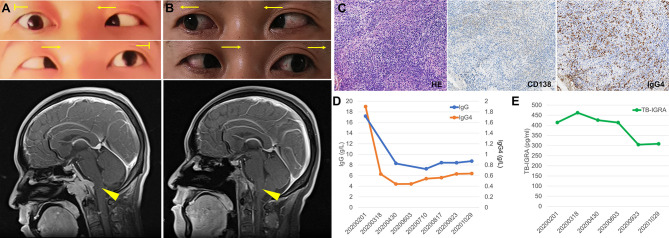
Figure 1.
Imaging, histologic and laboratory findings of the case. (A) The abduction of both eyes was limited (arrows indicate the direction of movement), and contrast-enhancing brain magnetic resonance imaging (MRI) showed a 3.8×2.9×2.9 cm mass overlying clivus with dural tail sign (arrow head). (B) Abduction of both eyes recovered after treatment and MRI showed the mass in the clivus area was remarkably smaller. (C) Histologic features of the lesion revealed fibro-connective tissue with mixed inflammation containing predominantly plasma cells and immunochemical analysis revealed an increased number of IgG4-positive plasma cells (×200). (D, E) Serum levels of IgG and IgG4 (D) and T cell-released INF-γ in TB-IGRA (E) of the patient during the follow-up.

