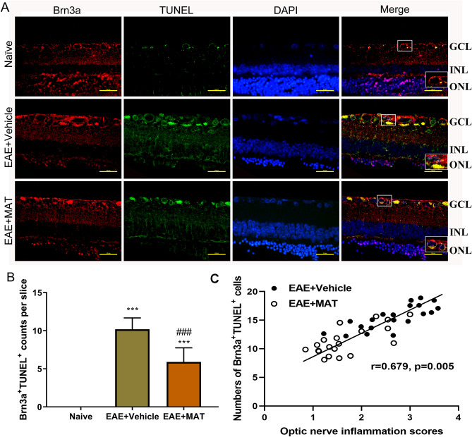
Figure 6.
MAT treatment protected RGCs from apoptosis. Neuroprotective effects of MAT were evaluated by counting RGCs immune-labeled with Brn3a antibody and estimating the number of RGC deaths through TUNEL. (A) RGCs in the both sides of temporal retina were examined by immunofluorescent double staining by anti-Brn3a (red) and TUNEL (green), and all cells were co-stained with DAPI (blue). ONL, outer nuclear layer; INL: inner nuclear layer; GCL, ganglion cell layer. Scale bars = 100 µm. (B) Quantitative analysis for the numbers of apoptotic RGCs (TUNEL+ Brn3a+DAPI+). (C) Scatter plots between optic nerve inflammation scores and numbers of Brn3a+TUNEL+ cells showing significant positive correlation. Numbers of Brn3a + TUNEL + cells were compared between EAE + Vehicle and EAE + MAT groups using generalized estimating equation (GEE) models with optic nerve inflammation scores as a covariate to adjust for within-subject inter-eye correlations. Subsequently, an adjusted P value was used for multiple comparison according to the Bonferroni correction methods. ***P < 0.001, comparison with the naive group. ###P < 0.001, comparisons between vehicle- and MAT-treated groups.

