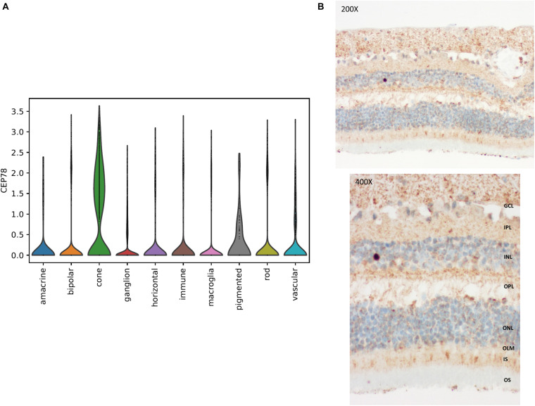
FIGURE 5.
Single-cell transcriptional data in human retina and immunohistochemistry of CEP78 in adult human retina. (A) Violin plot for visualization of scaled and corrected expression of CEP78 in 19,768 human neural retinal cells. Cells belonging to the cone cell population exhibit the highest expression. (B) Immunostaining of the CEP78 protein is visible in light brown. Light microscopy at 200 × (top) and 400 × (bottom). Predominant cytoplasmic expression of CEP78 can be seen in both cone and rod photoreceptors with strong staining at the base of the inner segments. Antibody: LN2004459 (1:100, rabbit, LabNed). GCL, ganglion cell layer; IPL, inner plexiform layer; INL, inner nuclear layer; OPL, outer plexiform layer; ONL, outer nuclear layer; OLM, outer limiting membrane; IS, inner segments; OS, outer segments.

