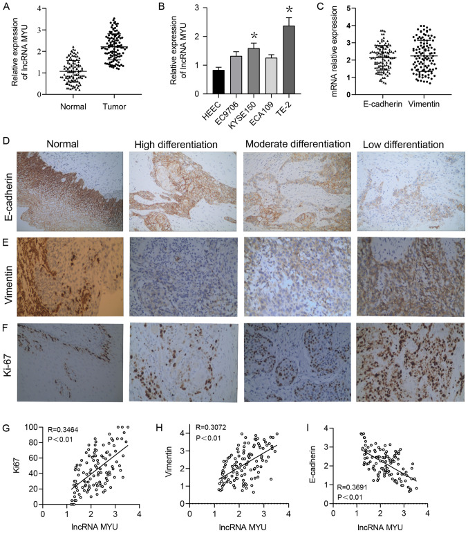
Figure 1.
Expression of lncRNA MYU, E-cadherin, Vimentin and Ki-67 in ESCC tissues. (A) The relative expression of lncRNA MYU was increased in ESCC tissues compared with that in normal tissues. GAPDH was used as a loading control. (B) Reverse transcription-quantitative PCR analysis of lncRNA MYU expression in 3 ESCC cell lines and HEECs. The relative expression level of lncRNA MYU was significantly upregulated in TE-2 cells. *P<0.05 vs. HEEC. (C) Relative mRNA expression of E-cadherin and Vimentin in ESCC tissues. GAPDH was used as a loading control. (D-F) Representative images of immunohistochemical staining for (D) E-cadherin, (E) Vimentin and (F) Ki-67 in ESCC tissues (magnification, x200). (G-I) Pearson correlation analysis of correlations between the relative expression of lncRNA MYU and that of (G) Ki-67, (H) Vimentin and (I) E-cadherin in ESCC tissues (P<0.01). ESCC, esophageal squamous cell carcinoma; HEECs, human esophageal epithelial cells; lncRNA, long non-coding RNA; MYU, c-Myc upregulated lncRNA.

