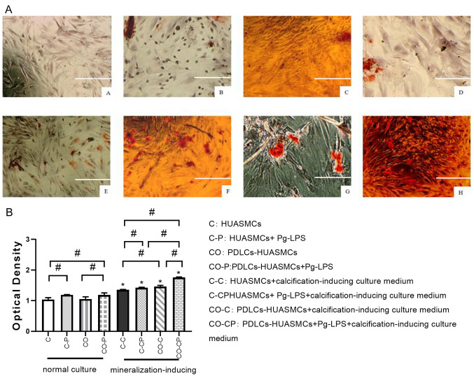
Figure 6.
Effect of Pg-LPS on calcified nodule formation of HUASMCs in different groups. (AA) HUASMCs (group C); (AB) HUASMCs + Pg-LPS (group C-P); (AC) PDLCs-HUASMCs (group CO); (AD) PDLCs-HUASMCs + Pg-LPS (group CO-P); (AE) HUASMCs in calcification-inducing culture medium (group C-C); (AF) HUASMCs + Pg-LPS in calcification-inducing culture medium (group C-CP); (AG) PDLCs-HUASMCs in calcification-inducing culture medium (group CO-C); and (AH) PDLCs-HUASMCs + Pg-LPS in calcification-inducing culture medium (group CO-CP). The calcified nodules were scattered in the C-P and CO-P groups and were almost absent in the C and CO groups. All of the groups under calcification induction or calcification induction + Pg-LPS (1 µg/ml) conditions exhibited many calcified nodules, whilst the co-cultured CO-C and CO-CP groups had slightly more calcified nodules than those cultured in DMEM medium (scale bar, 100 μm). The quantified results of calcification based on alizarin red S staining three weeks after calcification induction (B). The calcified nodule formation was significantly higher in calcification-inducing medium and co-culture groups treated with Pg-LPS (1 μg/ml) than in the control groups. Values are expressed as the mean ± standard deviation (n=3). #P<0.05, calcification-inducing culture medium vs. normal culture medium (i.e. group C-C vs. group C; group C-CP vs. group C-P; group CO-C vs. group CO; group CO-CP vs. group CO-P); *P<0.05. HUASMCs, human umbilical artery smooth muscle cells; PDLCs, periodontal ligament cells; Pg-LPS, Porphyromonas gingivalis lipopolysaccharide..

