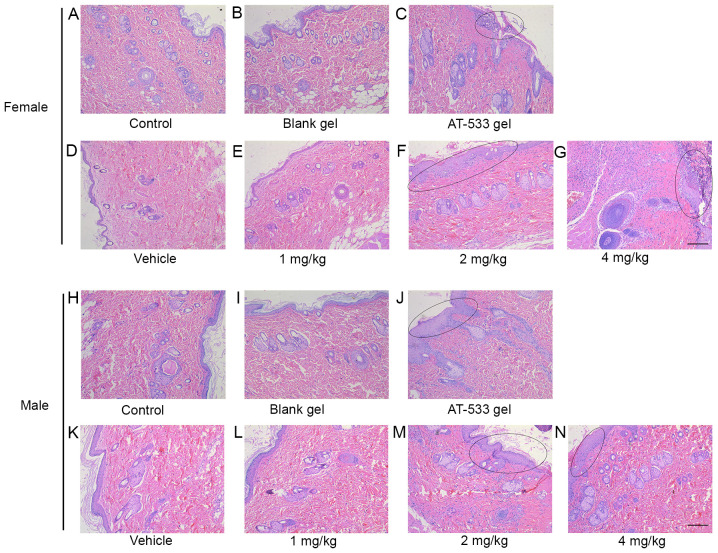
Figure 3.
Histological analysis of rat skin treated with AT-533 or AT-533 gel in the 30-day subacute toxicity. Representative micrographs of skin sections from female rats under the conditions of (A) control, (B) blank, (C) AT-533 gel, (D) vehicle, (E) 1 mg/kg AT-533, (F) 2 mg/kg AT-533 and (G) 4 mg/kg AT-533 stained with hematoxylin and eosin. Representative micrographs of skin sections from male rats under the conditions of (H) control, (I) blank, (J) AT-533 gel, (K) vehicle, (L) 1 mg/kg AT-533, (M) 2 mg/kg AT-533 and (N) 4 mg/kg AT-533 hematoxylin and eosin. The elliptical circle represents epidermal keratinization, thickening and inflammatory cell infiltration. Magnification, x10.

