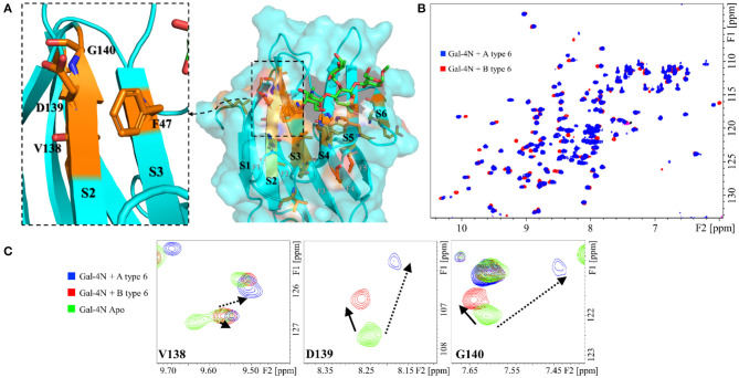
Figure 4.
Protein backbone CSP analysis for the interaction of Gal-4N with A and B type-6 tetrasaccharide antigens. (A) Molecular model (MD simulation) for the complex of Gal-4N with A type-6 antigen. The residues that significantly differ in their chemical shift perturbation upon binding to group A or B antigens are highlighted in orange, as well as F47 (unknown resonance assignment). Left: zoom at the S2–S3 region showing residues F46, V138, D139, and G140. (B) Superimposition of the 1H-15N HSQC spectra of Gal-4N in the presence of A type-6 (blue) and B type-6 antigens (red). (C) Expansion of the 1H-15N HSQC spectra at residues V138, D139, and G140: in green, Gal-4N apo; in blue, Gal-4N/A type-6 (12 eq.); and in red Gal-4N/B type-6 (10 eq.).

