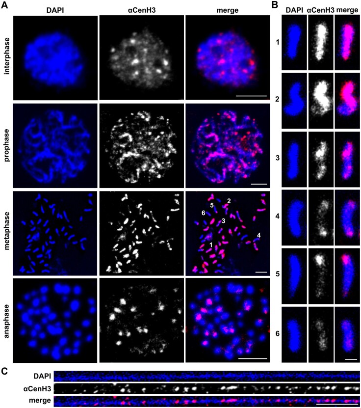
Fig. 4.
The organization of αCenH3 centromeres in Meloidogyne incognita. Slides were prepared from isolated reproductive tissue of females (ovaries and uterus). (A) Immunofluorescence of αCenH3-containing domains (red) during the mitosis cycle in M. incognita using anti-αCenH3 antibodies raised in rabbit 2 (supplementary fig. 3A, Supplementary Material online). Scale bar = 5 µm. (B) Distribution pattern of αCenH3-containing domains along metaphase chromosomes in six different chromosome types. Selected chromosome types were indicated in metaphase spread with numbers. Scale bar = 1 µm. (C) Immunofluorescence of αCenH3-containing domains (red) on chromatin fiber. Scale bar = 5 µm. All chromosomes and fibers were counterstained with DAPI (blue). Images were acquired with confocal microscopy and shown as z-stack projection.

