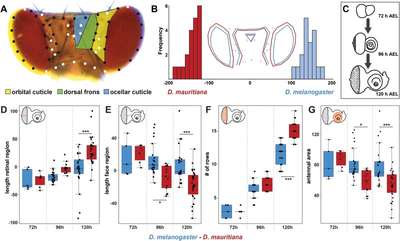
Fig. 1.
Quantitative differences in dorsal head shape are defined late during eye–antennal disc development. (A) Dorsal view of a Drosophila melanogaster head. The head cuticle consists of three morphologically distinguishable regions, namely the orbital cuticle (yellow) next to the compound eye, the dorsal frons (green), and the ocellary cuticle (blue). The 57 landmarks that were used to analyze head shape are shown as fixed (white) and sliding landmarks (black). (B) Discriminant function analysis distinguishes mean dorsal head shapes of D. melanogaster (blue) and D. mauritiana (red). Difference between means: Procrustes distance: 0.094, P value = 0.0001. (C) The development was characterized, and transcriptomic data sets were generated for developing eye–antennal discs for both species at three developmental stages: 72 h AEL (early L3), 96 h AEL (mid L3), and 120 h AEL (late L3). (D) Distance from the optic stalk to the morphogenetic furrow was measured along the equator region. Significant differences were observed at 120 h AEL (F5,96 = 15.61, P = 3.2e−11). (E) Distance from the morphogenetic furrow to the antennal anlagen was measured. From 96 h AEL on, we observed significant differences (F5,96 = 10.23, P = 7e−8). (F) Number of ommatidial precursor rows was counted along the equator region of the eye–antennal disc. Significant differences were observed at 120 h AEL (F5,96 = 210.8, P < 2e−16). (G) Area of the antennal region of the eye–antennal disc. Significant differences were observed at 96 and 120 h AEL (F5,96 = 7.86, P = 3.08e−6). One-way ANOVA followed by Tukey multiple comparisons: ***<0.001, *<0.05.

