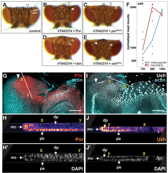
Fig. 4.
Pnr and Ush interact during eye–antennal disc development. (A) Dorsal view of an adult control head of D rosophila melanogaster (CyO carrying offspring of the VT042374 > Pnr cross that do not contain the UAS-Pnr transgene and thus no overexpression). Posterior (pVT) and anterior vertical bristles (aVT) are labeled. The arrowhead marks the occipital region. (B) Overexpression of pnr did not lead to major irregularities in the dorsal head cuticle, but to duplication of the pVT bristle next to the compound eye (white arrowhead) (9/9 females and 10/10 males showed the phenotype). (C) Knockdown of pnr resulted in visible enlargement of the occipital region (black arrowhead) and to the loss of the pVT bristle next to the compound eye (white circle) (10/10 males and females, respectively, showed the phenotype). (D) Overexpression of ush resulted in irregularities in the dorsal head cuticle and to the loss of the pVT andaVT bristles (white circles) next to the compound eye (10/10 females showed the phenotype, males were not tested). (E) Knock-down of ush resulted in slight irregularities of the dorsal head cuticle (11/11 females showed the phenotype, males were not tested), to loss (white circle) (10/10 females showed the phenotype, males were not tested) or misplacement (white arrowhead) of the aVT bristle (2/10 females showed the phenotype, males were not tested). (F) Expression dynamics of the ush transcript at the three developmental stages in D. melanogaster (blue) and D. mauritiana (red) based on rlog transformed read counts. (G, H) Pnr protein location in third instar eye–antennal discs in D. melanogaster. The Pnr protein is present in the dorsal peripodial epithelium (pe) of the developing disc (G), including a few cells of the margin cells (mc) and the disc proper (dp) (H′). The white arrow in G marks the morphogenetic furrow, the solid white line marks region of the cross-section shown in H and H′, and the x and y coordinates indicate the same location in G, H, and H′. (I, J) Ush protein location in third instar eye–antennal discs in D. melanogaster. The Ush protein is, similar to Pnr (compare with G), expressed in the dorsal peripodial epithelium (pe) of the developing disc (I), including a few cells of the margin cells (mc) and the disc proper (dp) (J′). The white arrow in I marks the morphogenetic furrow, the solid white line marks region of the cross-section shown in J and J′, and the x and y coordinates indicate the same location in I, J, and J′. G and I are maximum intensity projections of confocal sections throughout the eye–antennal disc. The scale bars represent 50 µm.

