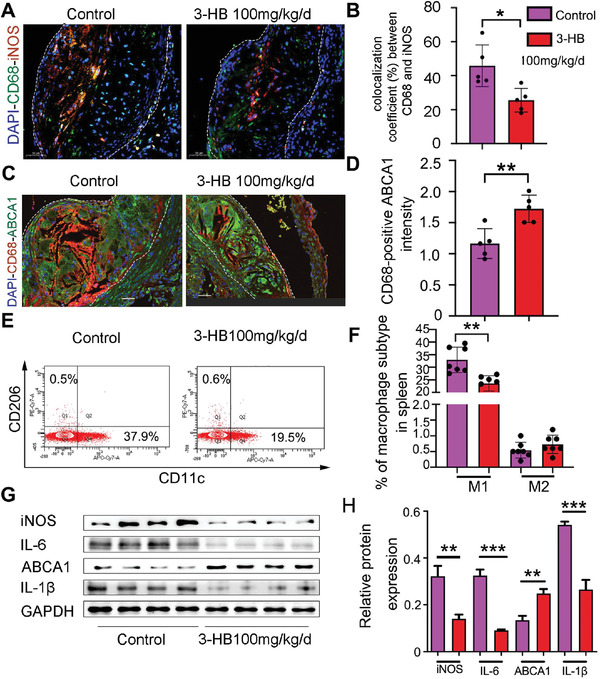
Figure 3.

3‐HB reduces M1 macrophages and promotes cholesterol efflux in apoE −/− mice. A,B) Immunofluorescence staining for iNOS (red) and CD68 (green) in the aortic root sections and quantification of colocalization, percentage of iNOS+ area in CD68+ area, respectively. DAPI was stained for cell nucleus (blue), Scale bar = 50 µm (n = 5 per group). C,D) ABCA1 expression (green) in macrophages (red) was assessed by immunofluorescence staining and quantification of ABCA1 fluorescence intensity overlapping with CD68+ area, respectively. Scale bar = 30 µm (n = 5 per group). E,F) Western blot analysis of inflammatory markers (IL‐6, iNOS, and IL‐1β) and cholesterol efflux marker (ABCA1) in the aortas from mice treated with or without 3‐HB and quantification of protein expression, respectively (n = 4 per group). G,H) Flow cytometry plots and quantitative analysis of macrophage differentiation in the spleen of apoE −/− mice treated with or without 3‐HB (100 mg kg−1 d−1) (n = 7 per group), respectively. Data are presented as the mean±SEM from at least three independent experiments, Student's t‐test, * p < 0.05, ** p < 0.01, and *** p < 0.001.
