
Figure 1. Heterogeneous exosome release from polarized MDCK cells.
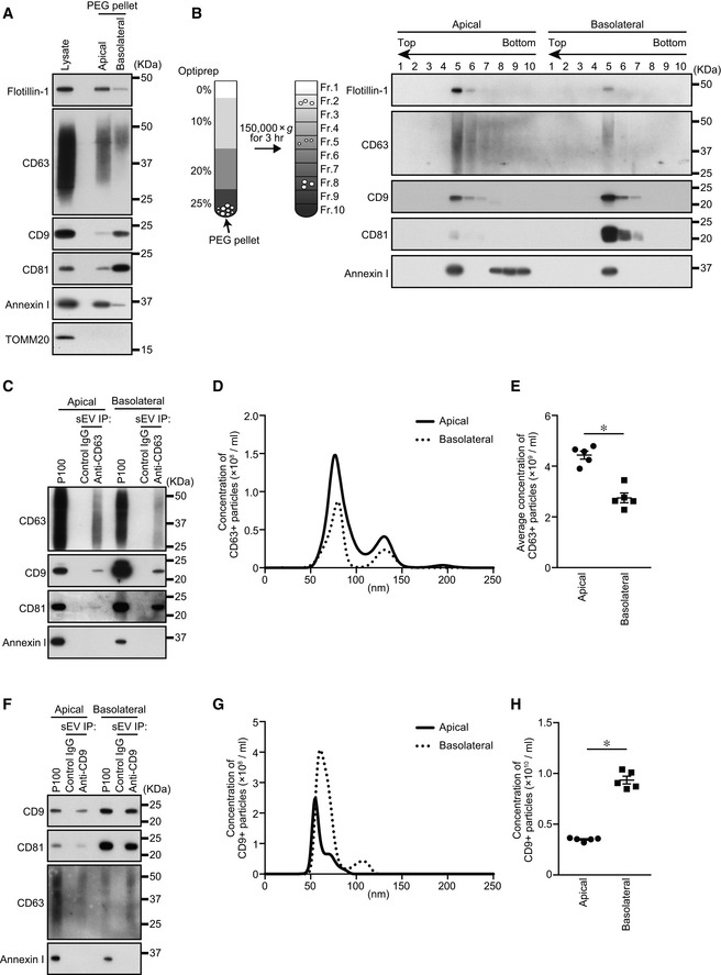
- MDCK cells were cultured on cell culture inserts for 4 days. On the last day, the culture medium was replaced with EV‐depleted medium. EVs released from the apical and basolateral sides of MDCK cells were purified by PEG precipitation. Cell lysates and EV proteins in PEG pellets were analyzed by immunoblotting with the antibodies indicated. Note that the PEG pellets did not contain mitochondrial protein TOMM20, suggesting that the PEG pellets were not contaminated by intracellular organelles.
- PEG pellets prepared as in (A) were subjected to OptiPrep flotation analysis.
- MDCK cells stably expressing human CD63 were cultured as in (A). sEVs were isolated from the pre‐cleared medium by direct immunoaffinity capture using anti‐CD63 antibody.
- sEVs prepared as in (C) were eluted from the beads with a glycine buffer and analyzed by nanoparticle tracking analysis (NTA). Representative NTA traces were shown.
- Quantification of the NTA data obtained in five independent experiments.
- MDCK cells were cultured as in (A). sEVs were isolated from the pre‐cleared medium by direct immunoaffinity capture using anti‐CD9 antibody.
- sEVs prepared as in (F) were eluted from the beads with a glycine buffer and analyzed by NTA. Representative NTA traces were shown.
- Quantification of the NTA data obtained in five independent experiments.
Data information: (A and B) Annexin I blots were separately obtained on different days using the same samples. (E and H) *P < 0.01 (two‐sided Student's unpaired t‐test). Mean ± s.e.m. was shown.