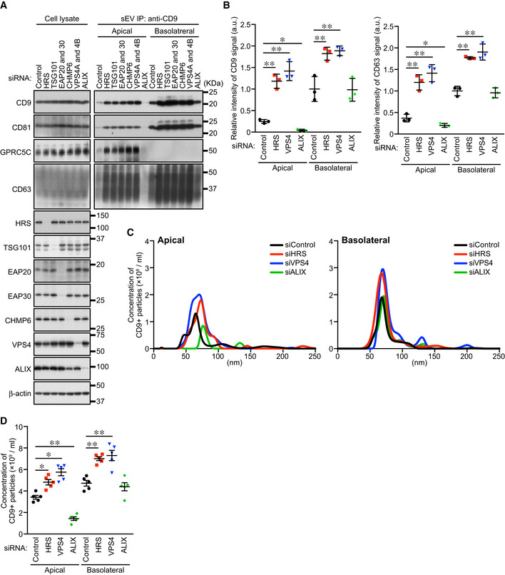
Figure 3. ALIX, but not the ESCRT machinery, is required for apical exosome release.

- MDCK cells were transfected with siControl or the siRNAs indicated, and the cells were transferred to cell culture inserts and cultured for 4 days. On the last day, the culture medium was replaced with EV‐depleted medium. sEVs were isolated from the pre‐cleared medium by direct immunoaffinity capture using anti‐CD9 antibody. Cell lysates and sEV samples were analyzed by immunoblotting with the antibodies indicated.
- The intensity of the bands shown in (A) was measured in three independent experiments.
- sEVs prepared as in (A) were eluted from the beads with a glycine buffer and analyzed by NTA. Representative NTA traces were shown.
- Quantification of the NTA data obtained in five independent experiments.
Data information: (A) CD63 blots were separately obtained on different days using the same samples. (B and D) *P < 0.05, **P < 0.01 (one‐way ANOVA and Tukey's test). Mean ± s.e.m. was shown.
