Figure 4.

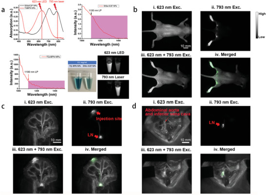
Dual‐channel NIR‐II fluorescence images of LNs and blood vessels. a) The differences of absorption and photoluminescence spectra between IDSe‐IC2F NPs and TQ‐BPN NPs, and the NIR‐II fluorescence images of IDSe‐IC2F NPs and TQ‐BPN NPs under the irradiation of 793 nm laser and 623 nm LED, respectively. b) i) Whole‐body blood vessel imaging of a mouse intravenously injected with TQ‐BPN NPs, under the excitation of 623 nm LED (before injection of IDSe‐IC2F NPs, 30 mW cm−2, exposure time (HG): 100 ms, 1100 nm LP); ii) sciatic and popliteal LNs imaging of the mouse treated with IDSe‐IC2F NPs, under the excitation of 793 nm laser (40 mW cm−2, HG: 100 ms, 1100 nm LP); iii) simultaneous imaging of whole‐body blood vessels and LNs, under the excitation of both 623 nm LED and 793 nm laser; iv) merged image of (ii) and (iii). Scale bar: 10 mm. c) i) Mesenteric vessels imaging of a rat receiving caudal vein injection of TQ‐BPN NPs, upon the excitation of 623 nm LED (before injection of IDSe‐IC2F NPs); ii) LNs imaging of the rat, which was injected with IDSe‐IC2F NPs from follicles and excited with the 793 nm laser; iii) simultaneous imaging of mesenteric vessels and LN, under the excitation of both 623 nm LED and 793 nm laser; iv) merged image of (ii) and (iii). Scale bar 10 mm; d) i) imaging of major blood vessels in retroperitoneal area of a rat intravenously injected with TQ‐BPN NPs (before injection of IDSe‐IC2F NPs, 623 nm LED excitation); ii) retroperitoneal LNs imaging of the rat treated with IDSe‐IC2F NPs (793 nm laser excitation); iii) simultaneous imaging of major blood vessels and LNs, under the excitation of both 623 nm LED and 793 nm laser; iv) merged image of (ii) and (iii). Scale bar: 10 mm.
