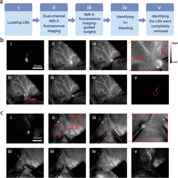
Figure 5.

Dual‐channel NIR‐II fluorescence imaging‐guided lymphadenectomy on rats. a) Brief description of the process. b) i) The precisely located mesenteric LNs labeled with IDSe‐IC2F NPs under 793 nm laser excitation; ii) dual‐channel fluorescence imaging of LNs and blood vessels (labeled with TQ‐BPN NPs) under the excitation of both 793 nm laser and 623 nm LED; iii) the process of resection (two LNs were detected and removed); iv) no obvious bleeding was observed; v) no residual LN was detected. Scale bar: 10 mm. c) i) The located gastric LNs labeled with IDSe‐IC2F NPs under 793 nm laser excitation; ii) dual‐channel fluorescence imaging of LNs and gastric vessels (labeled with TQ‐BPN NPs) under the excitation of both 793 nm laser and 623 nm LED; iii) the process of resection (the LN was very close to the left gastric artery); iv) no obvious bleeding was observed; v) no residual LN was detected. Scale bar: 10 mm. Red circles: the position of LNs (after resection).
