Figure 6.

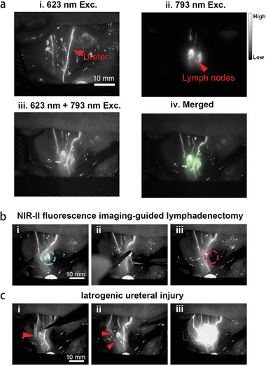
Dual‐channel NIR‐II fluorescence imaging of retroperitoneal LNs and ureters and imaging‐guided lymphadenectomy on rats. a) i) Retrograde ureteropyelography via TQ‐BPN NPs based NIR‐II fluorescence imaging (623 nm LED excitation, before the injection of IDSe‐IC2F NPs); ii) NIR‐II fluorescence image of retroperitoneal LNs labeled with IDSe‐IC2F NPs, under the excitation of 793 nm laser; iii) simultaneous imaging of ureters and LNs under the excitation of both 623 nm LED and 793 nm laser; iv) merged image of (ii) and (iii). Scale bar: 10 mm. b) The process of imaging‐guided lymphadenectomy (blue circle: the position of LNs, before resection): i) pulling the ureters aside; ii) resecting the LN; iii) confirming the LN was completely removed (red circle: the position of LNs, after resection). c) Simulative iatrogenic ureteral injury: i,ii) the broken end of ureter could be detected, red arrow: the broken end of ureter; iii) TQ‐BPN NPs leaked into abdominal cavity from the broken end of ureter as revealed by the fluorescence signal.
