
Figure 1.
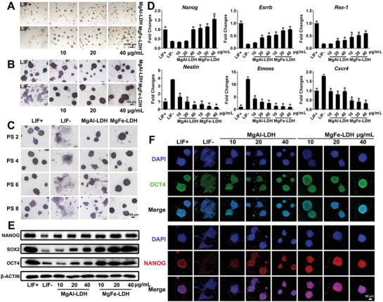
MgFe‐LDH is superior to MgAl‐LDH in supporting mESC self‐renewal. A) Morphology of clones observed via bright‐field microscopy. B) Representative images of mESCs cultured with nanoparticles and stained with ALP after 3 d of culture. C) ALP staining of mESCs treated with 20 µg mL−1 MgAl‐LDH or MgFe‐LDH at the indicated passage. D) qPCR analysis of key pluripotency genes (Nanog, Esrrb, and Rex‐1) and key differentiation genes (Nestin, Eomes, and Cxcr4). * represents p < 0.05, when compared to the LIF‐ treatment. E) Protein levels of NANOG, SOX2, and OCT4 in mESCs quantified by western blot. * represents p < 0.05, when compared to the LIF‐group. F) Representative confocal microscopy images of mESCs stained with OCT4 (green), NANOG (red), and DAPI (blue) accompanied by various treatments.