-
A
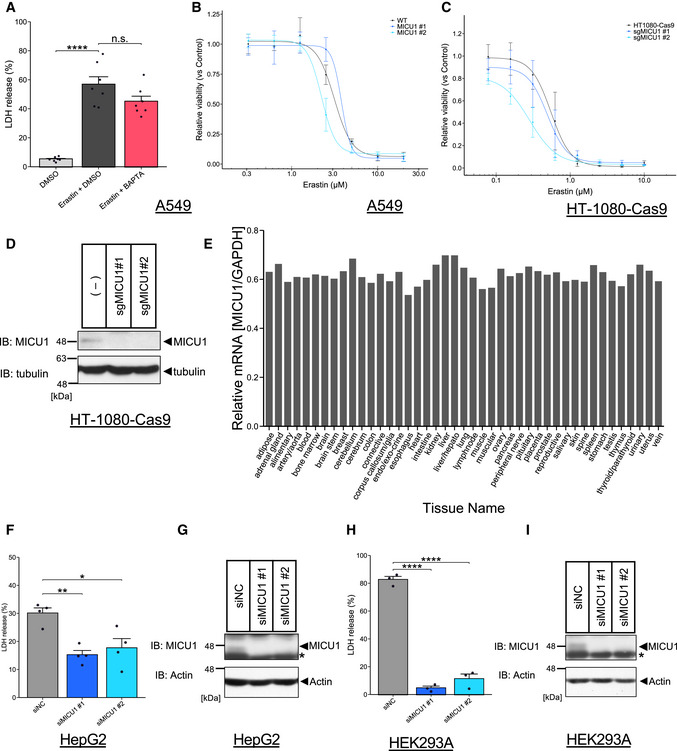
LDH release was measured after DMSO or erastin (10 µM) treatment with DMSO or BAPTA‐AM (10 µM) for 24 h. Data are presented as mean ± SEM; n = 7, biological replicates, ****P < 0.0001, n.s.: not significant, one‐way ANOVA followed by Bonferroni’s test.
-
B,C
Cell viability was measured after erastin treatment as indicated concentrations for 24 hours in A549 cells and MICU1 #1/#2 KO A549 cells or Cas9‐HT‐1080 cells and Cas9‐HT‐1080 cells infected with lentivirus containing sgRNA of MICU1 #1/#2. Data are presented as mean ± SEM; n = 3 biological replicates.
-
D
Immunoblots of MICU1 were present for knockout confirmation in Cas9‐HT‐1080 cells after lentivirus infection.
-
E
Relative mRNA expression of MICU1 in normal human tissue was normalized by GAPDH mRNA expression. Data was obtained from Refex database.
-
F
LDH release was measured after cold stress for 24 h in indicated cell lines. Data are presented as mean ± SEM; n = 4, biological replicates, **P < 0.01, *P < 0.05, one‐way ANOVA followed by Dunnett’s test.
-
G
Immunoblots of MICU1 were present for knockdown confirmation in HepG2 cells after siRNA transfection.
-
H
LDH release was measured after cold stress for 24 h in indicated cell lines. Data are presented as mean ± SEM; n = 3, biological replicates, ****P < 0.0001, one‐way ANOVA followed by Dunnett’s test.
-
I
Immunoblots of MICU1 were present for knockdown confirmation in HEK293A cells after siRNA transfection.

