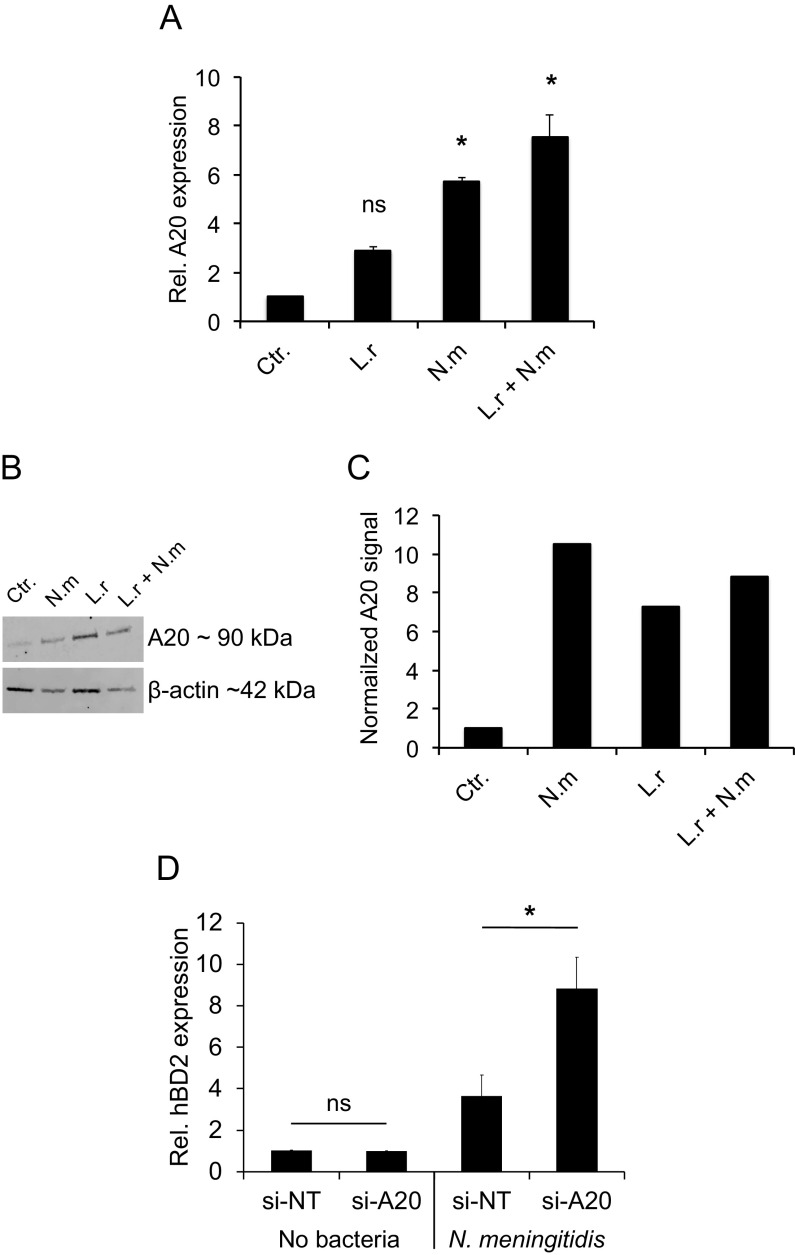
FIG 6.
N. meningitidis induces A20 expression. Pharyngeal epithelial cells were incubated with L. reuteri and N. meningitidis, either alone or coincubated, at an MOI of 100 for 6 h. (A) Gene expression of A20 was quantified using qPCR. Expression was normalized against the β-actin housekeeping gene and expressed as the fold change compared to the control. Data are represented as the mean values, with error bars representing the standard deviations. The assay was performed in triplicate at least three times. Significance was tested against the control. (B) A20 protein level in cell lysates as determined by Western blotting and compared to the housekeeping protein β-actin. (C) Normalized A20 protein level relative to the control. (D) Pharyngeal epithelial cells were transfected with control siRNA (si-NT) or siRNA targeted against A20 (si-A20) for 24 h. The cells were then infected with N. meningitidis at an MOI of 100 for 6 h. Gene expression of hBD2 was quantified using qPCR, normalized against the β-actin housekeeping gene, and expressed as the fold change compared to the control. Data are represented as the mean values, with error bars representing the standard deviations. The assays were performed in triplicate at least three times. Significance was tested against the control or as indicated. *, P < 0.05.

