FIG 1.
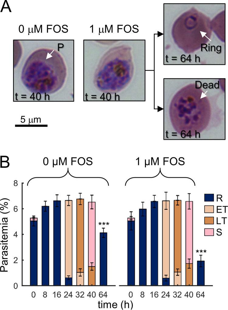
Morphologies and stages of the intraerythrocytic developmental cycle (IDC) of P. falciparum under untreated (0 μM) and fosmidomycin-treated (1 μM) conditions. (A) Giemsa-stained images of erythrocytes infected with P. falciparum (denoted by P) at 40 h into the first IDC. At 40 h, no gross morphological changes were apparent in parasite-infected erythrocytes treated with fosmidomycin (0 μM versus 1 μM fosmidomycin), of which ∼50% progressed to the ring stage of the second IDC (64 h, upper panel). (B) Percentage of ring-, (early and late) trophozoite-, and schizont-stage parasites in the infected cultures. Fosmidomycin treatment did not alter parasite development over time, as indicated by the fractions of different parasite stages before division, i.e., 40 h. During the second IDC in the absence of the drug, approximately half of the ring population was nonviable at the 64-h time point. At each time point, we collected parasitemia data in quadruplicate from independent experiments with the error bars indicating the standard deviation from the mean percentage for a given parasite stage. Asterisks denote a statistically significant (P ≤ 0.01) reduction in the ring population after treatment compared to an untreated control. Abbreviations: ET, early trophozoite; FOS, fosmidomycin; LT, late trophozoite; R, ring; S, schizont.

