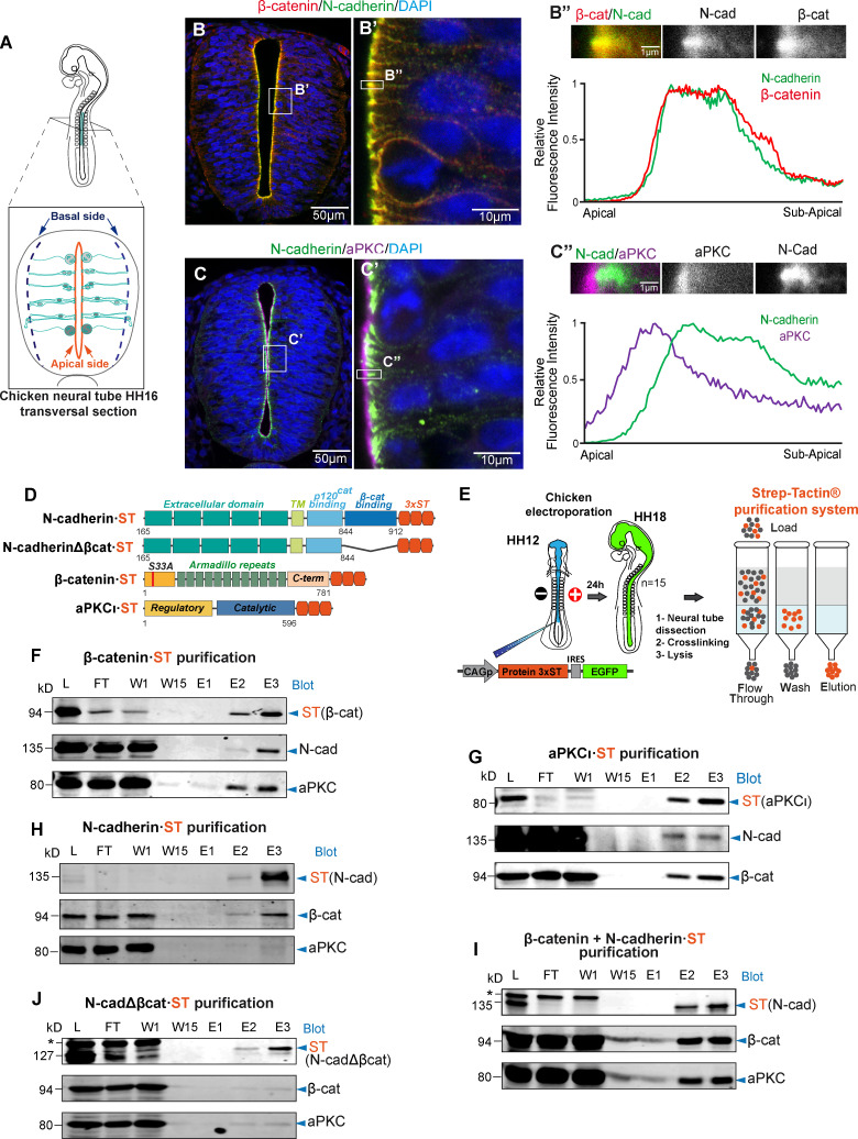
Figure 1.
β-catenin promotes the association of aPKCι with N-cadherin. (A) Representation of HH16 chicken NT slices. (B) Slices were stained with antibodies against β-catenin (red), N-cadherin (green), and DAPI (blue); the area labeled as B’ is enlarged in the right-hand panel. The graph shows the relative pixel intensity profile for N-cadherin and β-catenin in the area labeled as B’’, with the area quantified shown above. The two channels are displayed separately in grayscale for clarity. (C) As in B but using antibodies against N-cadherin and aPKCι. (D) Scheme of the four ST-tagged molecules used. (E) Representation of the in ovo electroporation and Strep-tag/Strep-Tactin purification procedures; 15 embryos (HH18 embryos) were used for each purification. (F–J) The different ST-tagged proteins were electroporated for 24 h into HH12 chicken NTs, and the ST-tagged and associated endogenous proteins were purified on Strep-Tactin columns at HH18. (F) Electroporation and purification of β-catenin·ST probed with antibodies against ST (β-catenin), N-cadherin, and PKCι. (G) Electroporation and purification of aPKCι·ST probed with antibodies against ST (aPKCι), N-cadherin, and β-catenin. (H) Electroporation and purification of N-cadherin·ST probed with antibodies against ST (N-cadherin), β-catenin, and aPKCι. (I) Electroporation of N-cadherin·ST plus untagged β-catenin. Purified proteins were probed with antibodies against ST (N-cadherin), β-catenin, and aPKCι. Note that electroporation of β-catenin substantially increased the amount of aPKC that copurified with N-cadherin·ST. (J) Electroporation and purification of N-cadherinΔβcat·ST probed with antibodies against ST (N-cadherinΔβcat), β-catenin, and PKCι. Note that neither β-catenin nor PKCι copurify with N-cadherinΔβcat·ST. In I and J, the asterisk indicates a nonspecific band. cad, cadherin; C-term, C-terminus; E, elution; FT, flow through; L, lysate; W, wash.

