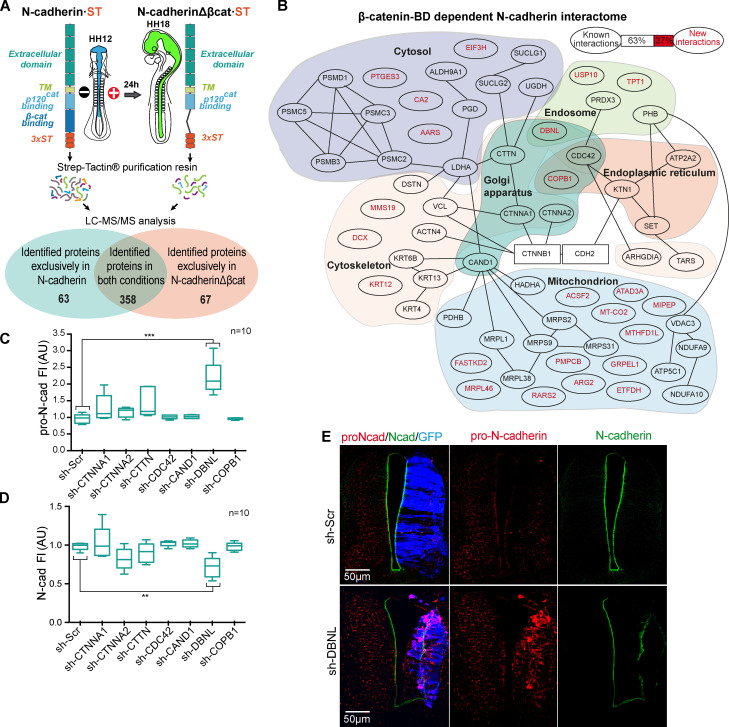
Figure 6.
Discovery of new β-catenin–dependent interactions with N-cadherin implicated in its maturation. (A) Scheme of the procedure followed to find new β-catenin–dependent interactions with N-cadherin. (B) The 63 proteins found in N-cadherin but not in N-cadherinΔβcat purification were assigned to subcellular compartments. The new (red) and known (black) interactions are shown, and known interactions are indicated by black lines in the tree. (C and D) Levels of pro-N-cadherin and N-cadherin, measured in HH23 chicken NTs transfected for 48 h with shRNAs targeting the proteins predicted to be located in the Golgi. Each experimental condition was compared with the respective control using a one-way ANOVA with a Dunnett’s multiple comparisons test (n = 10). (E) DBNL knockdown stained with antibodies against pro-N-cadherin (red) and N-cadherin (green). The GFP denoting transfection is shown in blue. **, P < 0.01; ***, P < 0.001. cad, cadherin; FI, fluorescence intensity; MS/MS, tandem mass spectrometry; Scr, scramble; TM, transmembrane domain; BD, binding domain.

