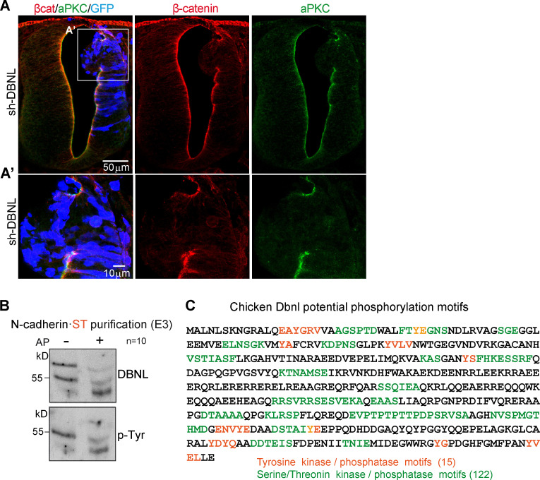
Figure S5.
DBNL knockdown disrupts the apico-basal polarity of chicken NT. (A and A’) HH23 chicken NTs were stained with β-catenin (red), aPKC (green), and GFP (blue, indicates transfection) 48 hpe with sh-DBNL. (B) Western blot of N-cadherin·ST–purified fractions untreated or treated with AP. The membrane was sequentially incubated with antibodies against Dbnl and phospho-Tyr. Note that the two Dbnl bands turned into a single band of lower molecular weight upon AP treatment, indicating that the difference between the two Dbnl bands was due to their distinct phosphorylation status. Note also that only the lower band was phosphorylated at Tyr (n = 10). (C) Scheme of the reported phosphorylation sites in chicken Dbnl as predicted with the PhosphoMotif finder tool (http://hprd.org/PhosphoMotif_finder); note that each individual Tyr, Ser or Thr may be part of different phosphorylation motifs.

