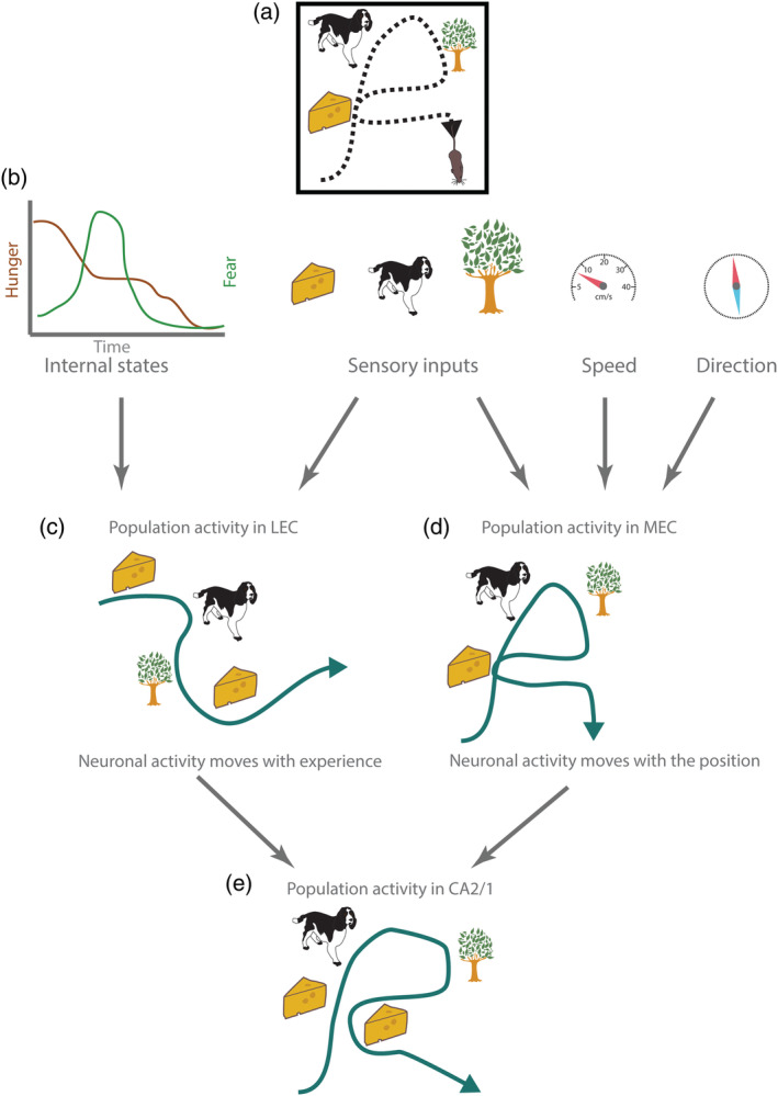
Figure 11.

Schematic overview of spatial and temporal codes in entorhinal cortex and hippocampus. (a) Agent moves and have four experiences (two of them in the same position). Black dashed line indicates path of the animal. (b) Information concerning internal states of the animal, sensory inputs, and self‐motion signals reach entorhinal cortex. (c) Neuronal population activity (green line) in lateral entorhinal cortex moves with the experience of the agent. (d) Neuronal population activity (green line) in medial entorhinal cortex moves with the position of the animal. (e) Neuronal population activity (green line) in CA2 and CA1 in hippocampus moves with the position and experience of the animal. Episodes are mapped in a spatiotemporal framework conveyed from entorhinal cortex. Neural activity is more similar (but not identical) during two visits to the same location [Color figure can be viewed at wileyonlinelibrary.com]
