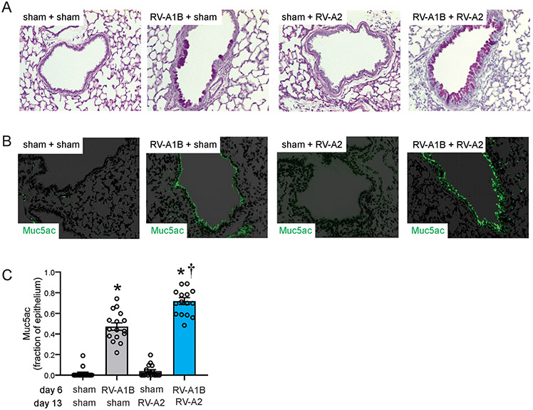
Figure 2. Heterologous RV infection induces exaggerated mucus metaplasia.
Baby mice were inoculated with sham or RV-A1B on day 6 and sham or RV-A2 on day 13 of life. Lungs were harvested on day 20 and processed for histology. Lungs sections were stained for PAS and Muc5ac and quantified using NIH ImageJ. A) PAS staining in sham, RV-A1B, RV-A2, RV-A1B + RV-A2-infected mice. The black bar is 50 microns (μ). B) Muc5ac staining in RV-A1B, RV-A2, RV1B + RV-A2-infected mice. The white bar is 50 μ. C) Quantification of Muc5ac staining in the airways. Data are represented are Muc5ac-positive cells per micron of basement membrane length. Images were taken at ×200 magnification. Data shown are mean ± SEM; n=3-4 airways/mouse, 4 mice per group from two different experiments; *different from sham + sham, †different from RV-A1B + sham, P < 0.05 by one-way ANOVA and Tukey multiple comparison test.

