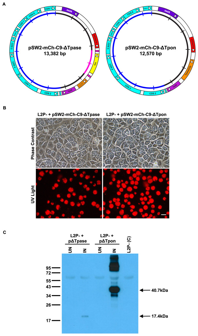
Figure 5. Design and functional analysis of the individual transposon and transposase vectors.
A: Plasmid maps showing the individual C. trachomatis/E. coli shuttle vectors pSW2-mCh-C9-ΔTpase and pSW2-mCh-C9-ΔTpon carrying either the transposon or the C9 transposase respectively. The shared sequence coloured in the outer circle shows the key CDS for chlamydial plasmid replication and maintenance (blue), spectinomycin selection (purple) and the mCherry marker (red) for visualisation of inclusions. In pSW2-mCh-C9-ΔTpase the active site of the C9 transposase has been deleted, in pSW2-mCh-C9-ΔTpon the entire transposon unit has been deleted. The inner circle is a scale with 1kb increments, the transposon sequence in this circle is coloured pink. B: Red fluorescent inclusions in McCoy cells infected by C. trachomatis L2P- transformed with either pSW2-mCh-C9-ΔTpase or pSW2-mCh-C9-ΔTpon vectors and grown under spectinomycin selection. Phase contrast and UV light images are from the same field at 48h.p.i. Left-hand panels are from L2P- containing pSW2-mCh-C9-ΔTpase and right-hand panels are from L2P- containing pSW2-mCh-C9-ΔTpon. Scale bar represents 20μm. C: Western Blot showing the induction on the C9 transposase in C. trachomatis L2P- transformed with either pSW2-mCh-C9-ΔTpase or pSW2-mCh-C9-ΔTpon vectors. Equal amounts of protein loaded in each gel lane. McCoy cells were infected with C. trachomatis L2P- transformed with either the pSW2-mCh-C9-ΔTpase or pSW2-mCh-C9-ΔTpon vectors. They were induced with ATc at 24h.p.i. and harvested at 48h.p.i. Non-induced and un-transformed L2P- were used as controls. Samples were separated by 12% SDS-PAGE gel and the presence of the truncated C9 transposase (arrowed at 17.4kDa) or C9 transposase (arrowed at 40.7kDa) was detected by Western blot with polyclonal antisera to purified C9 transposase.

