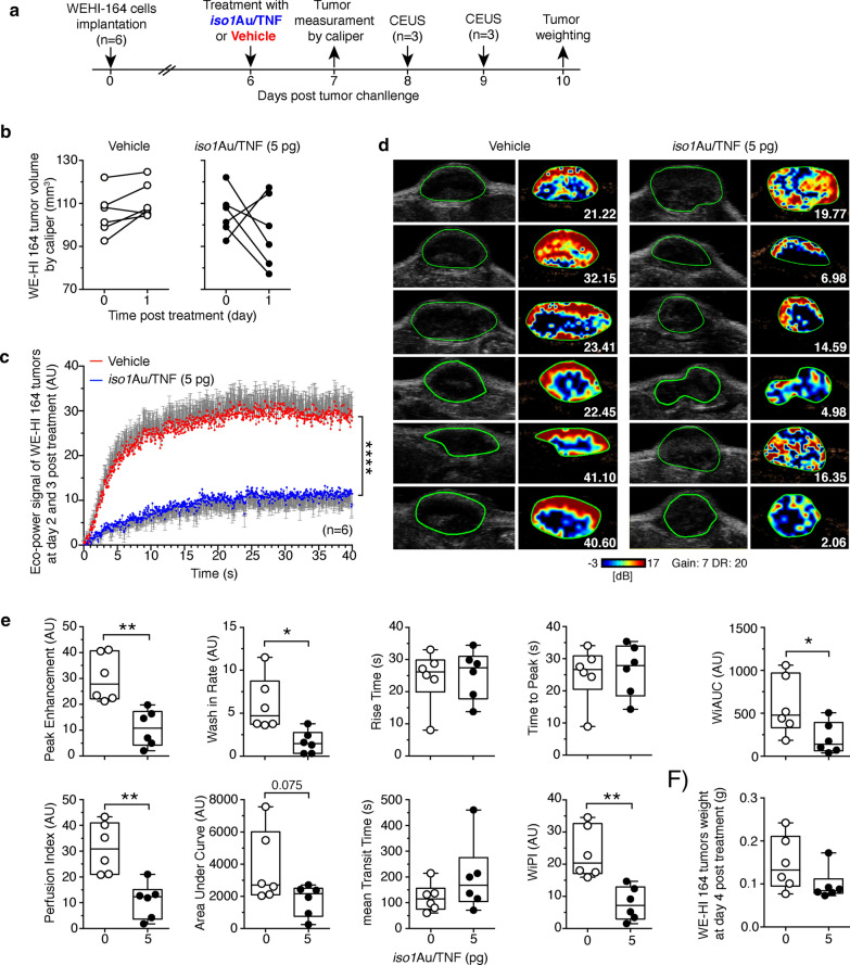
Fig. 6.
Iso1Au/TNF reduces tumor perfusion. WEHI 164 tumor-bearing mice (n = 6/group) were treated i.v. with the indicated dose of iso1Au/TNF at day 6 after tumor implantation and were analyzed by contrast-enhanced ultrasound (CEUS) at day 8 (3 mice) and day 9 (3 mice). MicroMarker Contrast Agent was injected at time 0, and its uptake was recorded for 40 s. a Scheme of the experiment. b Effect of iso1Au/TNF (5 pg) or vehicle on tumor growth before and after treatment (day 1), as determined by calipers. c Time-intensity curves (mean ± SE); ****p < 0.0001, by two-way ANOVA. d Grayscale tumor images (left) and color-coded peak enhancement (PE) maps of representative mice. Red and blue areas correspond to high and low perfused tumor areas, respectively. Numbers, PE values. e Quantitative analysis of tumor perfusion with microbubbles performed using VevoCQTM software. CEUS parameters were calculated using a region of interest (ROI, green lines in d) corresponding to the entire tumor area. Box plots of CEUS parameters with 5–95 percentiles and medians are shown; *P < 0.05; **P < 0.001 by two-tailed t-test. Images were taken at regions with a maximum tumor diameter. f Tumor weight at day 10

