. 2021 Apr 29;3(2):e200564. doi: 10.1148/ryct.2021200564

Figure 11:

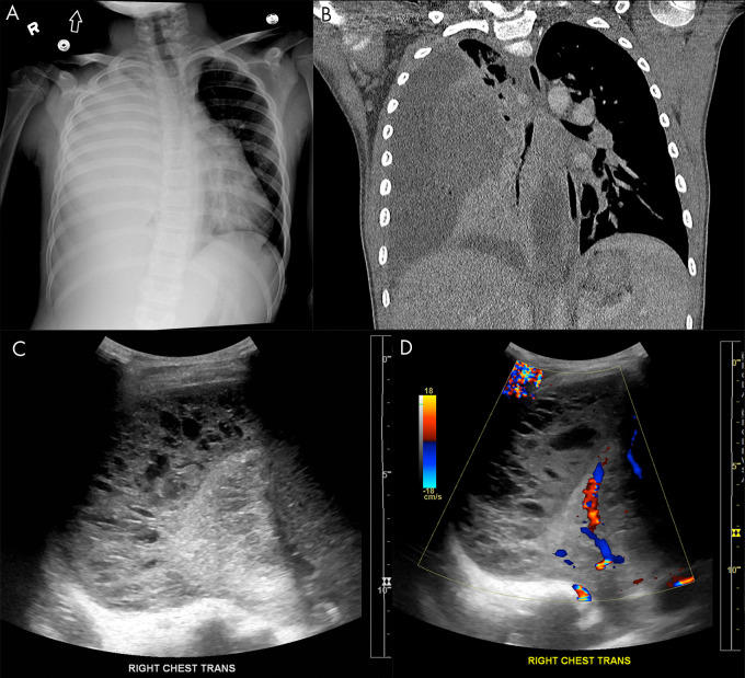
Empyema on lung US. A, Anteroposterior chest radiograph and B, coronal chest CT image show a large, right-sided empyema occupying the pleural space with associated compression of the pulmonary parenchyma in a 9-year-old boy. Lung US C, grayscale and D, Doppler images demonstrate a multiloculated complex-appearing fluid collection in the pleural space with septations and internal debris without internal color flow.

Empyema on lung US. A, Anteroposterior chest radiograph and B, coronal chest CT image show a large, right-sided empyema occupying the pleural space with associated compression of the pulmonary parenchyma in a 9-year-old boy. Lung US C, grayscale and D, Doppler images demonstrate a multiloculated complex-appearing fluid collection in the pleural space with septations and internal debris without internal color flow.