. 2021 Mar 25;12(17):5977–5993. doi: 10.1039/d1sc00165e

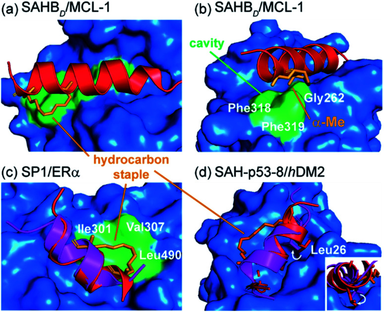
Fig. 4. Co-crystal structures of protein-bound constrained peptides highlighting the potential for contact between constraint and protein; (a) crystal structure of the MCL-1 SAHBD/MCL-1 complex (PDB: 3MK8)58 and the hydrocarbon staple (orange) of MCL-1 SAHBD makes additional hydrophobic contacts at the perimeter of the core interaction site; (b) a methyl group of the α,α-disubstituted functionality occupies a groove defined by Gly262, Phe318 and Phe319 of MCL-1 (green); (c) comparison of the ERα bound structures of the native sequence (purple, PDB: 2QGT) and the stapled peptide SP1 (red, PDB: 2YJD);60 (d) comparison of the hDM2 bound structures of the native p53 peptide (purple, PDB: 1YCR)61 and the stapled peptide (red, PDB: 3V3B).62.

Fig. 4