. 2021 Apr 6;12(17):6037–6047. doi: 10.1039/d0sc05328g

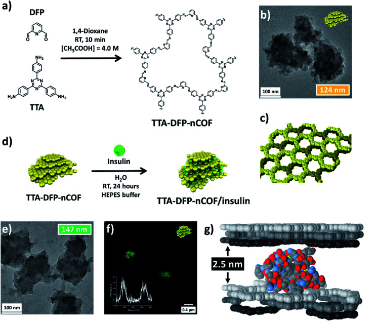
Fig. 1. Insulin is intercalated between TTA-DFP-nCOF layers. (a) Chemical structure and synthesis of the TTA-DFP-nCOF. (b) HR-TEM image of the TTA-DFP-nCOF. Cartoon representation (yellow nanoparticles) of the shape of the TTA-DFP-nCOF. (c) Structural model of the TTA-DFP-nCOF, consisting of hcb layers that are disposed in the abc sequence, generating hexagonal channels along the stacking direction. (d) Schematic representation of the encapsulation of insulin between the layers of the TTA-DFP-nCOF. Cartoon representation (green spheres) represents the insulin. (e) HR-TEM image of TTA-DFP-nCOF/insulin. (f) Confocal microscopy image of TTA-DFP-nCOF/insulin-FITC; inset: fluorescence intensity. (g) van der Waals representation of the optimized location of the insulin monomer molecule intercalated between TTA-DFP-nCOF layers. Atoms belonging to the COF layers are displayed in white, grey, and black color, for each individual layer. For the insulin molecule, the C, N, O, and S atoms are grey, blue, red, and yellow, respectively. H atoms are omitted for clarity.

Fig. 1