
FIG. 1.
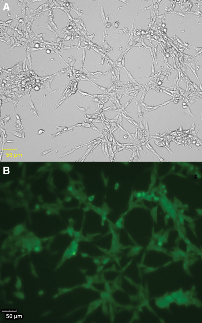
Imaging of ROS in neurons. Live cell imaging of N27 rat dopaminergic neurons treated with 250 μM of ammonium iron (III) citrate for 3 h. (A) Bright-field imaging. (B) DCFDA fluorescence as an indicator of ROS in cells. DCFDA, 2′,7′-dichlorofluorescin diacetate; ROS, reactive oxygen species. Color images are available online.