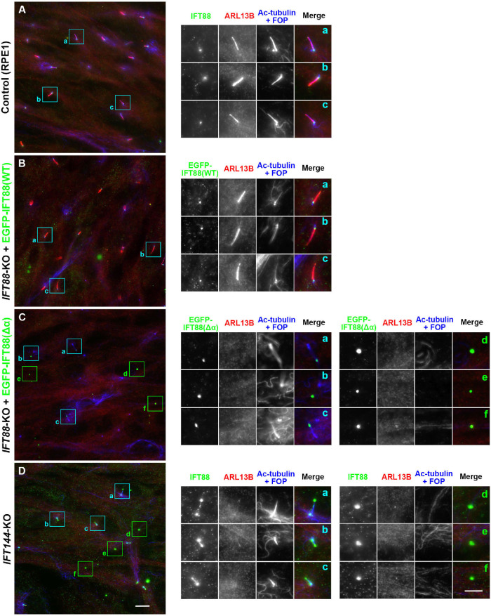
FIGURE 5:
Presence of ECVs formed by IFT88(Δα)-expressing IFT88-KO cells and by IFT144-KO cells. Control RPE1 cells (A), IFT88-KO cells expressing EGFP-IFT88(WT) (B) or EGFP-IFT88(Δα) (C), or IFT144-KO cells (D) were serum-starved for 24 h to induce ciliogenesis and immunostained for IFT88, ARL13B, and Ac-tubulin+FOP. Scale bar, 10 µm. Images of the boxed regions enlarged 2.5 times are shown in a–f. Scale bar, 5 µm.

