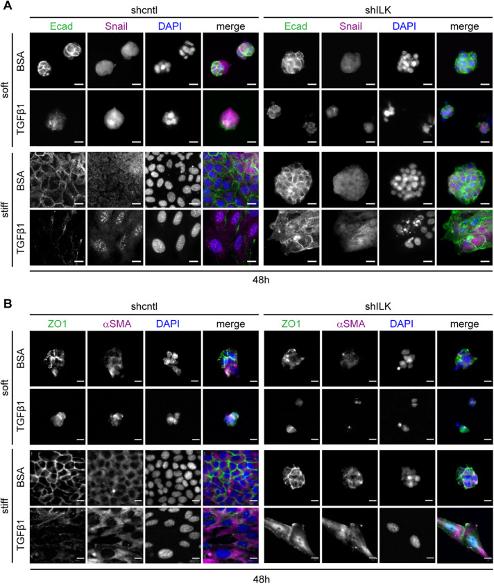
FIGURE 3:
On stiff substrata, depletion of ILK leads to either cell detachment or a delayed EMT after prolonged exposure to TGFβ1. Immunofluorescence analysis for (A) E-cadherin (green), Snail (magenta), and nuclei (blue), or (Β) ZO1 (green), αSMA (magenta), and nuclei (blue) in shcntl and shILK cells cultured on soft or stiff substrata and treated with or without TGFβ1 for 48 h. Scale bars, 10 μm.

