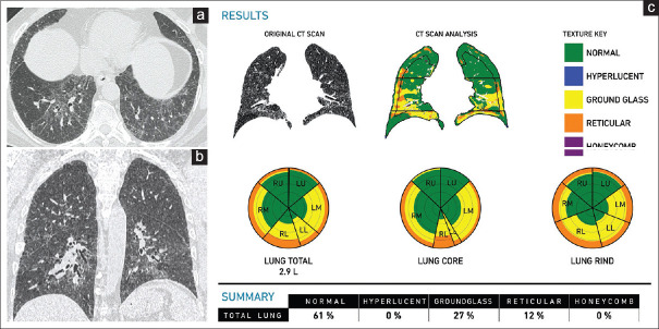
Figure 5.
(a-c) Chronic hypersensitivity pneumonitis. A 57-year-old man. Axial (a) and coronal. (b) Computed tomography scan images show ground-glass attenuation with reticular opacities and axial distribution, findings consistent with the clinical impression of chronic hypersensitivity pneumonitis. The Computer-Aided Lung Informatics for Pathology Evaluation and Rating analysis (c) shows total lung involvement of 39% with ground glass of 27% and a pulmonary vessel-related structure score of 5.2%

