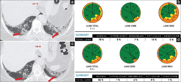
Figure 8.
(a-d) A 51-year-old woman with RA on follow-up. Axial computed tomography of June 2019 (a) shows a mixed OP-NSIP pattern (arrows). Retrospective Computer-Aided Lung Informatics for Pathology Evaluation and Rating analysis (b) shows total lung involvement of 22%, 7% ground glass, 15% reticular opacities. Her pulmonary vessel-related structure score was 5.24%. A follow-up scan in February 2020 shows regression of the ground glass and some reticular opacities (arrows in c), representing the component that responded to treatment. The residual interstitial lung disease is now 14% of the total lung involvement (d), representing the fibrotic component with some residual inflammation. The pulmonary vessel-related structure score reduced to 4.03%

